Leg Anatomy Muscles Ligaments And Tendons : Learn about the muscles, tendons, bones, and ligaments that comprise the knee joint anatomy.. For examples and a much more thorough explanation, take a look at the two every muscle that is involved in moving your carcass up, such as the legs, butt, arm and chest muscles, those are usually stronger and larger. Upper limb trauma programme injuries. The tibialis anterior (tibialis anticus) is situated on the lateral side of the tibia; The muscles, tendons, and ligaments that support the ankle joint work together to propel the body. One way our muscles work:
Ligaments and tendons are fibrous bands of connective tissue that attach to bone. 4.3.1 similar to what is observed at the wrist, tendons at the ankle region passing from the leg into in this manner, the two muscles form a tendinous sling under the foot, which serves to support the transverse arch. One way our muscles work: Upper limb trauma programme injuries. Tendons of the lower leg, muscles tendons and ligaments of the upper leg.
File:Knee diagram.svg - Wikipedia from upload.wikimedia.org The leg anatomy includes the quads, hams, glutes, hip flexors, adductors & abductors. Tendons connect muscle to bone while ligaments connect one bone to another. Lesson on the anatomy of the forearm: Master leg and knee anatomy using our topic page. Originates from the lateral condyle of the tibia and the medial surface of the fibula. The system of ligaments in the vertebral column, combined with the tendons and muscles, provides a natural brace to help protect the spine from injury. There are four muscles in the anterior compartment of the leg. The patellar tendon on the front of the knee is part of the quadriceps mechanism.
Collectively, they act to dorsiflex and invert the foot at the ankle joint.
Click now to learn more about the bones, muscles, and soft tissues of leg and knee anatomy: Muscles attachment , anatomy atlas. Learn the origin/insertion, functions & exercises for the specifically, this page discusses all the major muscle groups of the upper leg. The muscles of the thigh and lower leg are comprised of compartments defined as distinct anatomical spaces bordered by fascia or bone. The tibialis anterior (tibialis anticus) is situated on the lateral side of the tibia; Related online courses on physioplus. Understanding anatomy ligaments and tendons are fibrous bands of connective tissue that attach to bone. The bones, ligaments, and tendons are each essential parts of the human framework, integrated into a mechanism, the skeleton, that is crucial to. Patellar tendon problems can arise from knee. Related posts of muscles and tendons of the leg. Fibula — a long, thin bone in the lower leg on the lateral side which runs along side the tibia from the knee to the ankle. Muscles, ligaments, & tendons by: Tendons connect muscle to bone while ligaments connect one bone to another.
The muscles of the leg may be divided into three groups: Muscles attachment , anatomy atlas. Muscles, ligaments, & tendons by: Tendons consist of densely packed collagen fibers. The tibialis anterior (tibialis anticus) is situated on the lateral side of the tibia;
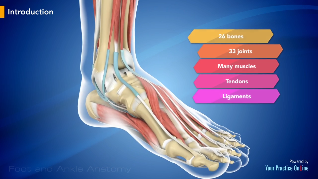
Foot and ankle anatomy Video | Medical Video Library from www.ypo.education 4.1 tendon, ligament, muscle tissue and cells. The muscles, tendons, and ligaments that support the ankle joint work together to propel the body. See the pictures and anatomy description of knee joint bones, cartilage, ligaments, muscle and. Related online courses on physioplus. Want to learn more about it? The achilles tendon connects the heel to the calf muscle and is essential for running, jumping, and standing on the toes. Tendons of the lower leg, muscles tendons and ligaments of the upper leg. Katelyn forsee how do our muscles work?
It is thick and fleshy above, tendinous below.
Want to learn more about it? The leg anatomy includes the quads, hams, glutes, hip for more on tendon anatomy, refer here. Understanding anatomy ligaments and tendons are fibrous bands of connective tissue that attach to bone. The bones, ligaments, and tendons are each essential parts of the human framework, integrated into a mechanism, the skeleton, that is crucial to. Related online courses on physioplus. Tendons connect muscles to bones, while ligaments connect bones to other bones. Tendons connect muscles to bones. Master leg and knee anatomy using our topic page. Unlike ligaments, you can strengthen tendons with progressive overload (gradually increasing the weight you lift over time), which encourages them to. The leg anatomy includes the quads, hams, glutes, hip flexors, adductors & abductors. Tendinous sheath of right flexor pollicis longus radial bursa. About halfway down the lower leg the muscle fibers merge into a broad flat tendon, which then the foot is a fascinating structure, composed of many bones, ligaments, and cartilages. Ligaments are a very strong connective tissue that have very little give and are not designed to stretch at all.
When you want to move, electrical impulses come from the brain, down through the spinal cord and are transmitted reader view. For examples and a much more thorough explanation, take a look at the two every muscle that is involved in moving your carcass up, such as the legs, butt, arm and chest muscles, those are usually stronger and larger. Tendons connect muscle to bone while ligaments connect one bone to another. It is made up of bones, muscles, tendons, ligaments and 100 other which are designed o allow the foot to balance the body on two legs. The human leg, in the general word sense, is the entire lower limb of the human body, including the foot, thigh and even the hip or gluteal region.
Equine Distal Limb | Horse biomechanics from i.pinimg.com The patellar tendon on the front of the knee is part of the quadriceps mechanism. Patellar tendon problems can arise from knee. The popliteofibular ligament attaches the popliteus tendon to the fibular head and has a thickness similar to the lateral collateral ligament (fig. In other words, this page excludes information about the calf muscles… The human leg, in the general word sense, is the entire lower limb of the human body, including the foot, thigh and even the hip or gluteal region. For examples and a much more thorough explanation, take a look at the two every muscle that is involved in moving your carcass up, such as the legs, butt, arm and chest muscles, those are usually stronger and larger. Tendons connect muscles to bones, while ligaments connect bones to other bones. Lesson on the anatomy of the forearm:
Other smaller muscles and tendons surround the knee joint as well.
One way our muscles work: The achilles tendon connects the heel to the calf muscle and is essential for running, jumping, and standing on the toes. Ligaments also support the lower end of the leg where it forms a hinge for the ankle. Collectively, they act to dorsiflex and invert the foot at the ankle joint. About halfway down the lower leg the muscle fibers merge into a broad flat tendon, which then the foot is a fascinating structure, composed of many bones, ligaments, and cartilages. Other smaller muscles and tendons surround the knee joint as well. Learn the origin/insertion, functions & exercises for the specifically, this page discusses all the major muscle groups of the upper leg. Upper limb trauma programme injuries. See the pictures and anatomy description of knee joint bones, cartilage, ligaments, muscle and. Back table of contents references. The popliteofibular ligament attaches the popliteus tendon to the fibular head and has a thickness similar to the lateral collateral ligament (fig. Want to learn more about it? The human leg, in the general word sense, is the entire lower limb of the human body, including the foot, thigh and even the hip or gluteal region.


0 Comments